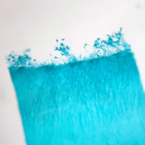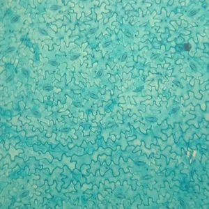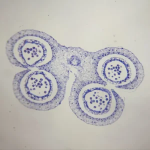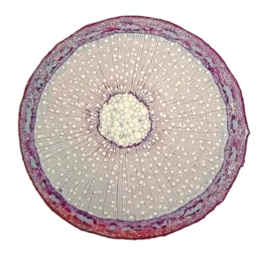
Skeletal Study Standard: Ranunculus Root, T.S. | 100% Dicot Vascular Architecture
$1.00
Ranunculus root prepared slide, Buttercup root cross section specimen
Dicot root anatomy histology, primary xylem microscopy slides, botany teaching specimens
Skeletal Study Standard: Ranunculus Root, T.S. | 100% Dicot Vascular Architecture
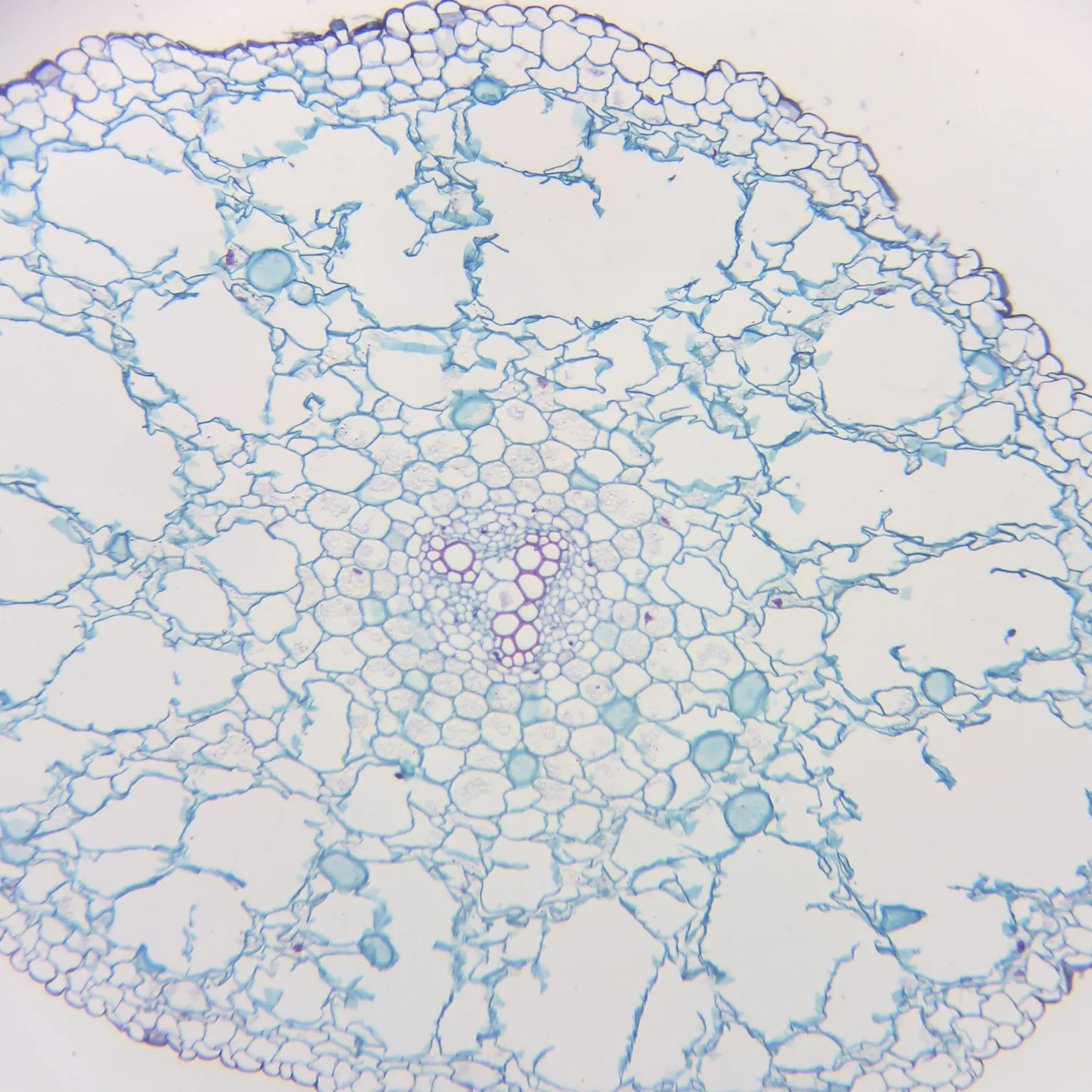
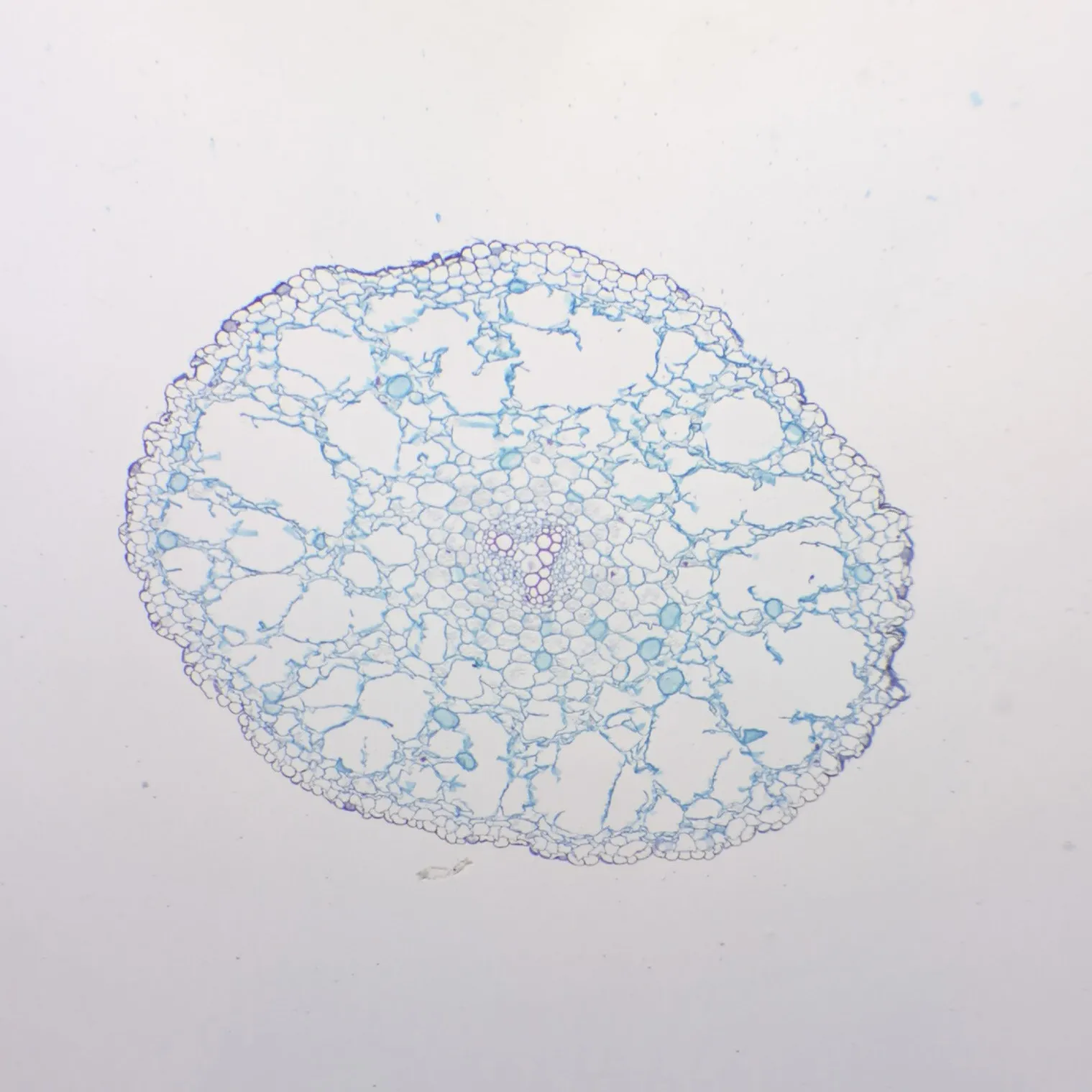
This Ranunculus root (Buttercup) transverse section (T.S.) slide provides an industry-standard, classic look at primary dicotyledonous root structures. Known globally as the #1 model organism for teaching plant anatomy, this Prepared Microscope Slide offers 100% clear visualization of uncorrupted tissue layers and a pristine central vascular cylinder.
Product Specifications & Quality Excellence:
-
Standard Dimensions: 25.4mm x 76.2mm (1″ x 3″) premium high-transparency glass slide.
-
Specimen Type: High-quality transverse cross section (T.S.) of a mature Ranunculus root.
-
Precision Quad-Contrast Staining: Expertly stained to deliver 100% sharp color differentiation between structural cell walls, starch-storing parenchyma, and lignified transport vessels.
-
Flawless Processing: Cut at an ultra-thin micrometer level to avoid tissue folding, ensuring every cell layer stays perfectly flat and intact for microscopic scanning.
4 Critical Botanical Highlights:
-
The Tetrarch/Triarch Core: Observe the #1 defining feature—the central star-shaped arrangement of primary xylem arms (displaying classic triarch or tetrarch symmetry) nested cleanly within patches of primary phloem.
-
Starch-Rich Cortex: Identify the massive cortical region packed with dense 100% distinct parenchyma cells, which are highly adapted for structural support and nutrient storage.
-
Crisp Endodermis Ring: View the specialized single-cell layer outlining the central cylinder, featuring distinct Casparian strips that regulate 100% of water filtration entering the vascular system.
-
Epidermis & Root Hair Base: Easily trace the protective outermost layer, presenting pristine cell boundaries that lack a cuticle to facilitate optimal absorption.
Botanical & Educational Significance:
-
The Ultimate Dicot Model: Serves as the 100% authoritative baseline reference for textbooks and laboratory workbooks across secondary and university-level botany courses.
-
Comparative Tissue Studies: An indispensable #1 tool for teaching students how to visually contrast a typical dicot root structure against monocot configurations (e.g., Zea mays or Dendrobium).
-
Vascular Differentiation Practice: Perfect for introducing beginners to xylem and phloem micro-anatomy under medium to high-power magnification (100X to 400X).
Reference & Further Reading:
For a comprehensive look, visit: Ranunculus – Wikipedia
Other fixed kits:
- 100pcs/set, https://www.ihappysci.com/product/100pcs-botany-prepared-microscope-slides/
- 25pcs/set, https://www.ihappysci.com/product/phanerogamae-prepared-microscope-slides/
Payment & Ordering
Ordering from us is simple and secure. We offer flexible payment options to suit your needs:
-
Online Orders: We primarily accept PayPal (Credit Card payments are supported via PayPal).
-
Alternative Methods: For high-volume orders or regional preferences, we also support Bank Transfer (T/T), Western Union, MoneyGram, WeChat Pay, and Alipay.
-
How to Order: Simply browse our catalog and checkout online, or [Contact Us] for a proforma invoice if using alternative payment methods.

Shipping & Global Delivery
We provide reliable worldwide shipping to ensure your laboratory supplies arrive safely and on time:
-
Fast Processing: Standard orders are processed and shipped within 3 business days.
-
Global Couriers: We partner with leading carriers, including DHL, FedEx, UPS, and official Postal services to offer both express and economy options.
-
Real-time Updates: If your order requires extra preparation time, our team will proactively contact you with a confirmed shipping schedule and tracking details.

Customization & OEM Services
Can’t find a specific specimen? As a leading manufacturer of prepared microscope slides, we specialize in bespoke solutions:
-
Custom Quotations: Send us your specific list, and our technical team will provide a detailed quote within 12 hours.
-
OEM & Private Label: We offer professional OEM services to create customized slide sets tailored to your curriculum or branding requirements.
-
Bulk Inquiries: From rare biological sections to large-scale educational sets, we have the expertise to deliver exactly what you need.
Key Terminology for Microscope Slides
To help you select the most suitable specimen for your research or teaching, please refer to our standard sectioning abbreviations:
-
W.M. (Whole Mount): The entire organism or structure is preserved and mounted on the slide.
-
C.S. (Cross Section): A thin transverse section through the organism or tissue plane.
-
L.S. (Longitudinal Section): A vertical section along the longest plane of the organism.
-
C.S. & L.S.: Both Cross and Longitudinal sections are mounted on the same slide for side-by-side comparison.
Extensive Catalog: Over 3,000+ Prepared Slides
As a leading Microscope Prepared Slides Manufacturer from China, we provide a comprehensive range of biological specimens across multiple disciplines:
| Product Category | Variety Count | Product Category | Variety Count |
| 🌿 Botany | 868 Kinds | 🧬 Cell Biology & Genetics | 64 Kinds |
| 🔬 Histology | 375 Kinds | 🧪 Botany Pathology | 373 Kinds |
| 🦁 Zoology | 459 Kinds | ➗ Mitosis & Meiosis | 66 Kinds |
| 🐾 Animal Pathology | 150 Kinds | 🐣 Embryology | 75 Kinds |
| 🧫 Microbiology | 100 Kinds | 🦟 Parasitology | 120 Kinds |
| 🪨 Petrology (Rock Sections) | 291 Kinds | 🛠️ Customized Sets | Available |
Looking for Custom Microscope Slides?
We specialize in customized preparation to meet specific laboratory or educational requirements.
If your required items are not listed in our standard catalog, we’ve got you covered!
-
Fast Quotation: Email us your specific list, and receive a formal quote within 12 hours.
-
Global Support: Our technical team is ready to assist with your scientific discoveries.
Contact Us Today:
📧 Email: [email protected]
🟢 WhatsApp: +86 18838161683
❓ For more information, please visit our [FAQ Page].
| Weight | 0.1 kg |
|---|---|
| Dimensions | 15 × 20 × 10 cm |