Master-Grade Helianthus leaf microscope slide, 100% Detail of 4 Dicot Tissue Layers
$1.00
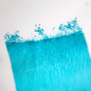
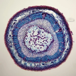
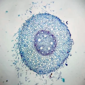
Helianthus leaf microscope slide, sunflower leaf C.S.
dicot leaf anatomy, plant histology specimens
mesophyll structure microscopy, botany educational slides
Master-Grade Helianthus leaf microscope slide, 100% Detail of 4 Dicot Tissue Layers
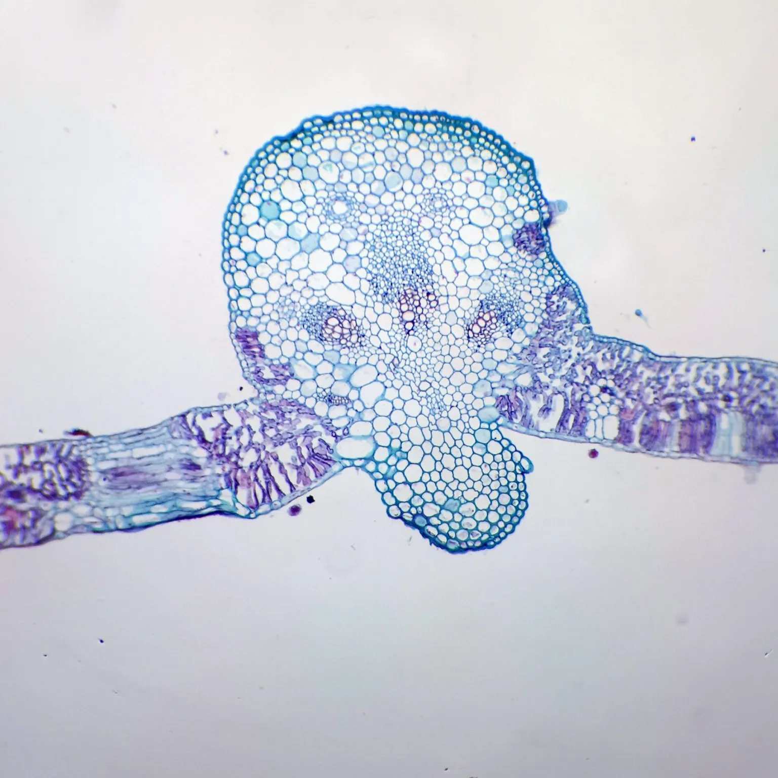
Our Helianthus Leaf microscope slides are expertly thin-sectioned to reveal the complex internal organization of a typical dicot leaf. This specimen is a 5-star choice for teaching plant anatomy, specifically the differentiation between palisade and spongy mesophyll.
Elite Product Specifications:
-
Dimensions: Standard 25.4mm x 76.2mm (1″ x 3″) microscope slide.
-
Staining: Professionally multi-stained to provide 100% color contrast between the vascular bundles (red/purple) and photosynthetic tissues (blue/green).
-
Magnification: Optimized for clear visualization at 40x (low power) and 100x-400x (high power).
4 Essential Histological Highlights:
-
Epidermis: Observe both the upper and lower epidermal layers, often featuring a protective waxy cuticle and specialized stomata for gas exchange.
-
Palisade Mesophyll: Densely packed, elongated cells located just below the upper epidermis, 100% optimized for maximum light absorption. * Spongy Mesophyll: Loosely arranged cells with large air spaces, facilitating the rapid diffusion of $CO_2$ and $O_2$ throughout the leaf.
-
Vascular Bundle (Vein): A prominent central midrib containing Xylem (for water transport) and Phloem (for sugar transport), typically surrounded by a protective bundle sheath.
Biological Significance:
-
Photosynthetic Efficiency: The distinct layering of the sunflower leaf showcases how plants maximize energy production while minimizing water loss.
-
Dicot Model: Helianthus is a premier model organism for studying the “reticulate” venation pattern characteristic of dicots.
-
Environmental Adaptation: The arrangement of tissues illustrates how leaves adapt to regulate transpiration in various light intensities.
Reference & Further Reading:
Source: Helianthus – Wikipedia
Other fixed kits:
- 100pcs/set, https://www.ihappysci.com/product/100pcs-botany-prepared-microscope-slides/
- 25pcs/set, https://www.ihappysci.com/product/phanerogamae-prepared-microscope-slides/
Payment & Ordering
Ordering from us is simple and secure. We offer flexible payment options to suit your needs:
-
Online Orders: We primarily accept PayPal (Credit Card payments are supported via PayPal).
-
Alternative Methods: For high-volume orders or regional preferences, we also support Bank Transfer (T/T), Western Union, MoneyGram, WeChat Pay, and Alipay.
-
How to Order: Simply browse our catalog and checkout online, or [Contact Us] for a proforma invoice if using alternative payment methods.

Shipping & Global Delivery
We provide reliable worldwide shipping to ensure your laboratory supplies arrive safely and on time:
-
Fast Processing: Standard orders are processed and shipped within 3 business days.
-
Global Couriers: We partner with leading carriers, including DHL, FedEx, UPS, and official Postal services to offer both express and economy options.
-
Real-time Updates: If your order requires extra preparation time, our team will proactively contact you with a confirmed shipping schedule and tracking details.

Customization & OEM Services
Can’t find a specific specimen? As a leading manufacturer of prepared microscope slides, we specialize in bespoke solutions:
-
Custom Quotations: Send us your specific list, and our technical team will provide a detailed quote within 12 hours.
-
OEM & Private Label: We offer professional OEM services to create customized slide sets tailored to your curriculum or branding requirements.
-
Bulk Inquiries: From rare biological sections to large-scale educational sets, we have the expertise to deliver exactly what you need.
Key Terminology for Microscope Slides
To help you select the most suitable specimen for your research or teaching, please refer to our standard sectioning abbreviations:
-
W.M. (Whole Mount): The entire organism or structure is preserved and mounted on the slide.
-
C.S. (Cross Section): A thin transverse section through the organism or tissue plane.
-
L.S. (Longitudinal Section): A vertical section along the longest plane of the organism.
-
C.S. & L.S.: Both Cross and Longitudinal sections are mounted on the same slide for side-by-side comparison.
Extensive Catalog: Over 3,000+ Prepared Slides
As a leading Microscope Prepared Slides Manufacturer from China, we provide a comprehensive range of biological specimens across multiple disciplines:
| Product Category | Variety Count | Product Category | Variety Count |
 Botany Botany |
868 Kinds |  Cell Biology & Genetics Cell Biology & Genetics |
64 Kinds |
 Histology Histology |
375 Kinds |  Botany Pathology Botany Pathology |
373 Kinds |
 Zoology Zoology |
459 Kinds |  Mitosis & Meiosis Mitosis & Meiosis |
66 Kinds |
 Animal Pathology Animal Pathology |
150 Kinds |  Embryology Embryology |
75 Kinds |
 Microbiology Microbiology |
100 Kinds |  Parasitology Parasitology |
120 Kinds |
 Petrology (Rock Sections) Petrology (Rock Sections) |
291 Kinds |  Customized Sets Customized Sets |
Available |
Looking for Custom Microscope Slides?
We specialize in customized preparation to meet specific laboratory or educational requirements.
If your required items are not listed in our standard catalog, we’ve got you covered!
-
Fast Quotation: Email us your specific list, and receive a formal quote within 12 hours.
-
Global Support: Our technical team is ready to assist with your scientific discoveries.
Contact Us Today:
 Email: [email protected]
 WhatsApp: +86 18838161683
 For more information, please visit our [FAQ Page].
| Weight | 0.1 kg |
|---|---|
| Dimensions | 15 × 20 × 10 cm |
Related products
-
 Botany Slides
Typical dicot stem, tilia stem c.s. prepared microscope slides wholesale
$1.00 Add to cart -
-
-