

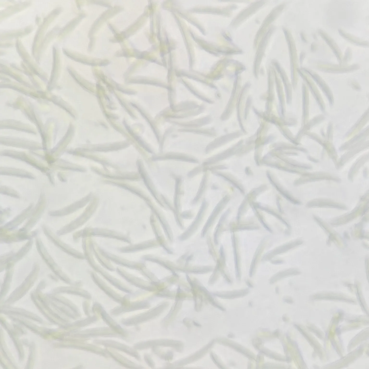
Gold Standard: Fusarium oxysporum Prepared Microscope Slide, 100% Spore Morphology & Vascular Wilt Study Accuracy
$3.00
Fusarium oxysporum Prepared Microscope Slide
Fusarium oxysporum prepared slide, Fusarium microscope whole mount
Phytopathology teaching specimens, vascular wilt fungi microscopy, macroconidia microconidia slide
Gold Standard: Fusarium oxysporum Prepared Microscope Slide, 100% Spore Morphology & Vascular Wilt Study Accuracy
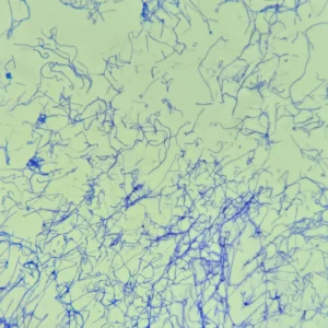
This professional whole mount (w.m.) slide provides a definitive visual standard for studying Fusarium oxysporum one of the world’s most devastating soil-borne plant pathogens. Famous for causing irreversible vascular wilt, root rot, and crown rot across a vast range of economically vital crops (including bananas, tomatoes, cotton, and legumes), this Prepared Microscope Slide offers 100% clear visualization of diagnostic microconidia, macroconidia, and associated hyphal networks.
The immaculate microscopic spread captured in Fusarium oxysporum highlights our advanced micro-dispersion preparation technique, presenting a highly uniform, high-density layer of individual spores without artificial clustering or cellular collapse, ensuring flawless identification under medium to high-power magnification.
Product Specifications & Quality Excellence:
-
Standard Dimensions: 25.4mm x 76.2mm (1″ x 3″) premium high-transparency glass slide.
-
Specimen Type: Expertly harvested whole mount (w.m.) capturing key reproductive macroconidia and vegetative mycelium structures.
-
Precision Contrast Staining: Professionally stained to deliver 100% high-definition contrast, accentuating the delicate septations, transparent cell walls, and curved geometries of the conidia against a clean optical background.
-
Archival Preservation: Bound with premium, non-yellowing mounting medium under a ultra-thin cover glass to protect the specimen from oxidation, degradation, and oil-immersion wear.
4 Critical Mycological & Phytopathological Highlights:
-
Sickle-Shaped Macroconidia: Observe the classic, highly recognizable diagnostic marker—slender, slightly curved or canoe-shaped macroconidia featuring thin walls, multi-septate partitioning, and a distinct pointed apical cell.
-
Abundant Oval Microconidia: Easily scan the abundant, small, single-celled or single-septate microconidia that dominate the vegetative phase, typically formed on short, unbranched conidiophores.
-
Septate Hyaline Mycelium: Trace the fine, branching, transparent (hyaline) septate hyphae that physically invade and plug the xylem vessels of host plants, causing systemic wilting.
-
Chlamydospore Evolution (Optional/In Culture): View the resilient, thick-walled resting spores (chlamydospores) formed within or at the tips of the hyphae, engineered for long-term soil survival.
Agricultural & Research Significance:
-
Phytopathology Reference Standard: Serves as a 100% authoritative baseline for agronomy faculties, plant pathology labs, and agricultural extension stations identifying vascular wilt agents.
-
Economic Crop Defense Education: An indispensable #1 visual aid for teaching the lifecycle of pathogens behind catastrophic agricultural crises, such as Panama Disease (Fusarium wilt) in bananas.
-
Taxonomic Differentiation Practice: Excellent for training students to visually isolate the delicate, curved macroconidia of Fusarium from the dark, partitioned muriform spores of Alternaria or the distosepta of Bipolaris.
Reference & Further Reading:
For a comprehensive look at: Fusarium oxysporum – Wikipedia
Other fixed kits:
- 100pcs/set, https://www.ihappysci.com/product/100pcs-botany-prepared-microscope-slides/
- 25pcs/set, https://www.ihappysci.com/product/phanerogamae-prepared-microscope-slides/
Payment & Ordering
Ordering from us is simple and secure. We offer flexible payment options to suit your needs:
-
Online Orders: We primarily accept PayPal (Credit Card payments are supported via PayPal).
-
Alternative Methods: For high-volume orders or regional preferences, we also support Bank Transfer (T/T), Western Union, MoneyGram, WeChat Pay, and Alipay.
-
How to Order: Simply browse our catalog and checkout online, or [Contact Us] for a proforma invoice if using alternative payment methods.

Shipping & Global Delivery
We provide reliable worldwide shipping to ensure your laboratory supplies arrive safely and on time:
-
Fast Processing: Standard orders are processed and shipped within 3 business days.
-
Global Couriers: We partner with leading carriers, including DHL, FedEx, UPS, and official Postal services to offer both express and economy options.
-
Real-time Updates: If your order requires extra preparation time, our team will proactively contact you with a confirmed shipping schedule and tracking details.

Customization & OEM Services
Can’t find a specific specimen? As a leading manufacturer of prepared microscope slides, we specialize in bespoke solutions:
-
Custom Quotations: Send us your specific list, and our technical team will provide a detailed quote within 12 hours.
-
OEM & Private Label: We offer professional OEM services to create customized slide sets tailored to your curriculum or branding requirements.
-
Bulk Inquiries: From rare biological sections to large-scale educational sets, we have the expertise to deliver exactly what you need.
Key Terminology for Microscope Slides
To help you select the most suitable specimen for your research or teaching, please refer to our standard sectioning abbreviations:
-
W.M. (Whole Mount): The entire organism or structure is preserved and mounted on the slide.
-
C.S. (Cross Section): A thin transverse section through the organism or tissue plane.
-
L.S. (Longitudinal Section): A vertical section along the longest plane of the organism.
-
C.S. & L.S.: Both Cross and Longitudinal sections are mounted on the same slide for side-by-side comparison.
Extensive Catalog: Over 3,000+ Prepared Slides
As a leading Microscope Prepared Slides Manufacturer from China, we provide a comprehensive range of biological specimens across multiple disciplines:
| Product Category | Variety Count | Product Category | Variety Count |
| 🌿 Botany | 868 Kinds | 🧬 Cell Biology & Genetics | 64 Kinds |
| 🔬 Histology | 375 Kinds | 🧪 Botany Pathology | 373 Kinds |
| 🦁 Zoology | 459 Kinds | ➗ Mitosis & Meiosis | 66 Kinds |
| 🐾 Animal Pathology | 150 Kinds | 🐣 Embryology | 75 Kinds |
| 🧫 Microbiology | 100 Kinds | 🦟 Parasitology | 120 Kinds |
| 🪨 Petrology (Rock Sections) | 291 Kinds | 🛠️ Customized Sets | Available |
Looking for Custom Microscope Slides?
We specialize in customized preparation to meet specific laboratory or educational requirements.
If your required items are not listed in our standard catalog, we’ve got you covered!
-
Fast Quotation: Email us your specific list, and receive a formal quote within 12 hours.
-
Global Support: Our technical team is ready to assist with your scientific discoveries.
Contact Us Today:
📧 Email: [email protected]
🟢 WhatsApp: +86 18838161683
❓ For more information, please visit our [FAQ Page].
| Weight | 0.1 kg |
|---|---|
| Dimensions | 15 × 20 × 10 cm |
Related products
-
-
-
Sale!
 Microbiology Slides
19pcs set of Fungi Prepared Microscope Slides, China Wholesale Supplier
Original price was: $50.00.$40.00Current price is: $40.00. Add to cart -
Sale!
 Microbiology Slides
Actinomycetes smear, bacteria prepared slides, expert wholesale supplier
Original price was: $3.00.$2.00Current price is: $2.00. Add to cart