Cup Fungus, Peziza Apothecium Sec., Prepared Microscope Slides
$2.00
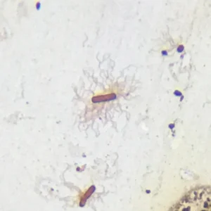
Cup Fungus, Peziza Apothecium Sec., Prepared Microscope Slides
Peziza microscope slide
Ascomycota sexual reproduction, ascus and ascospore histology
Cup fungus anatomy, mycology teaching slides
Cup Fungus, Peziza Apothecium Sec., Prepared Microscope Slides
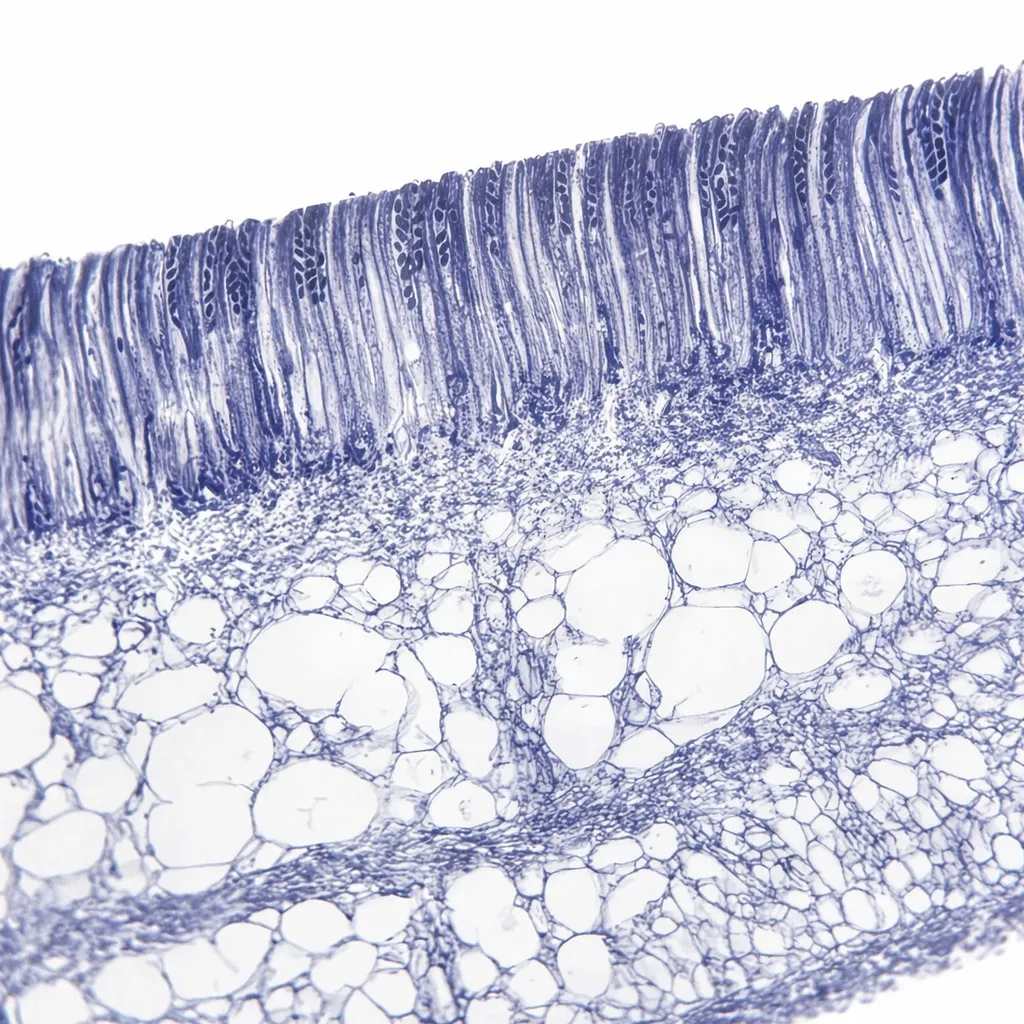
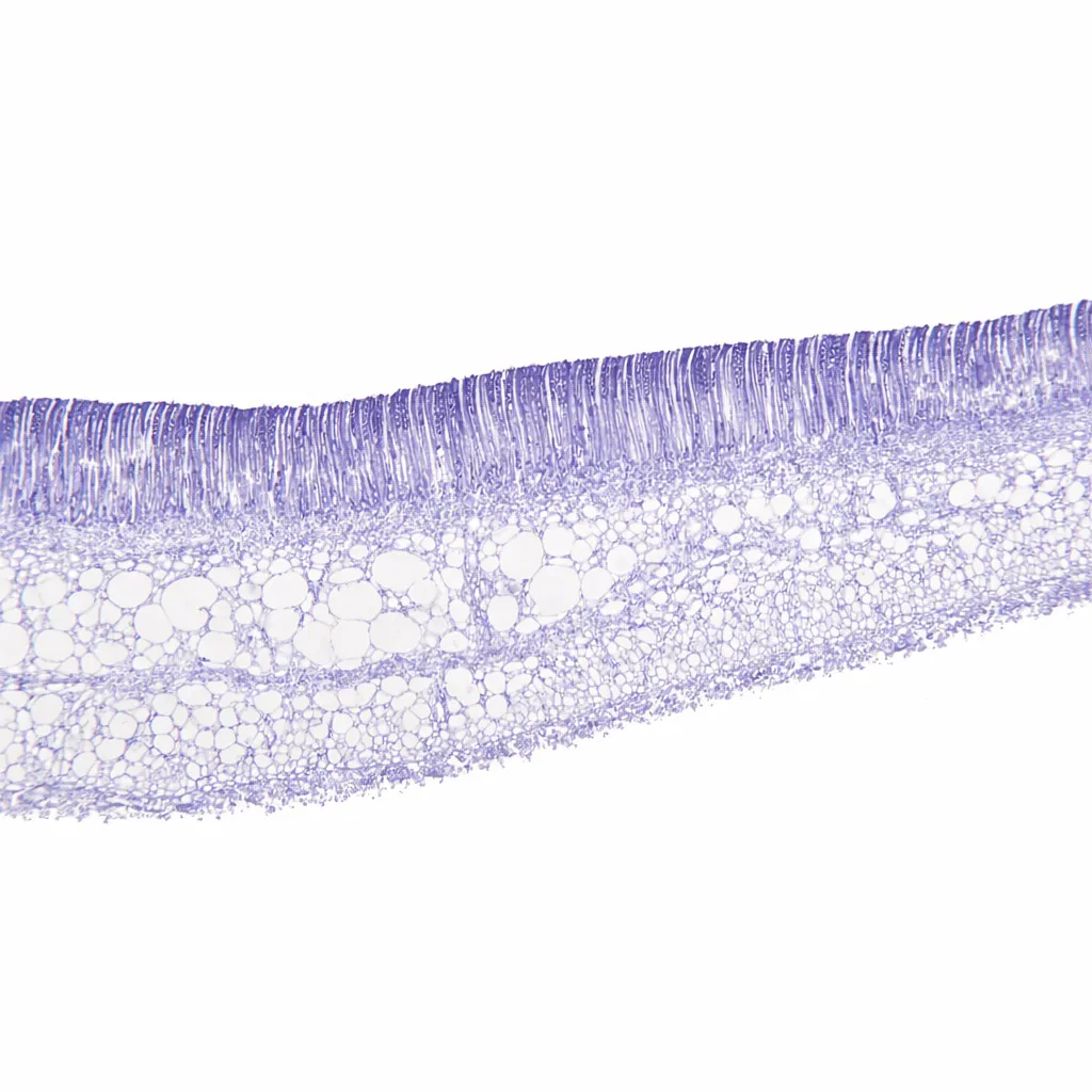
Our Peziza slides are expertly sectioned to show the highly organized layers of the fertile surface. This specimen provides a clear, high-contrast view of the microscopic “sac” structures that give Ascomycetes their name.
Product Specifications:
-
Dimensions: 25.4mm x 76.2mm (1″ x 3″)
-
Staining: Professionally stained with Cotton Blue or Hematoxylin. This specifically highlights the fungal chitin and the densely packed nuclei within the spores.
-
Section Type: Thin section (Sec.) through the apothecium to reveal the internal hymenial layer.
Key Mycological Features:
-
Hymenium Layer: The uppermost, fertile layer composed of vertically aligned asci and paraphyses.
-
Asci (Singular: Ascus): The elongated, sac-like cells. Each ascus typically contains eight ascospores, which are produced through meiosis followed by one mitotic division.
-
Ascospores: Observe the small, oval spores lined up inside each ascus. These are the primary means of sexual reproduction and dispersal.
-
Paraphyses: The sterile, hair-like filaments interspersed between the asci. These help support the asci and protect them until the spores are ready for discharge.
-
Subhymenium and Excipulum: The supportive fungal tissue (pseudoparenchyma) beneath the fertile layer that forms the body of the cup.
Biological Significance:
-
Sexual Reproduction: Peziza illustrates the definitive characteristic of the Ascomycota phylum: the production of spores within a sac-like ascus.
-
Spore Discharge: Many cup fungi use a pressurized mechanism to “puff” their spores into the air simultaneously, often triggered by changes in humidity or physical touch.
-
Ecological Role: Most Peziza species are saprobic, playing a vital role in the ecosystem by breaking down decaying organic matter like wood or soil.
Reference & Further Reading:
For a comprehensive look at the different forms of fungal fruiting bodies, visit: Peziza – Wikipedia
Other fixed kits:
- 100pcs/set, https://www.ihappysci.com/product/100pcs-botany-prepared-microscope-slides/
- 25pcs/set, https://www.ihappysci.com/product/phanerogamae-prepared-microscope-slides/
Payment & Ordering
Ordering from us is simple and secure. We offer flexible payment options to suit your needs:
-
Online Orders: We primarily accept PayPal (Credit Card payments are supported via PayPal).
-
Alternative Methods: For high-volume orders or regional preferences, we also support Bank Transfer (T/T), Western Union, MoneyGram, WeChat Pay, and Alipay.
-
How to Order: Simply browse our catalog and checkout online, or [Contact Us] for a proforma invoice if using alternative payment methods.

Shipping & Global Delivery
We provide reliable worldwide shipping to ensure your laboratory supplies arrive safely and on time:
-
Fast Processing: Standard orders are processed and shipped within 3 business days.
-
Global Couriers: We partner with leading carriers, including DHL, FedEx, UPS, and official Postal services to offer both express and economy options.
-
Real-time Updates: If your order requires extra preparation time, our team will proactively contact you with a confirmed shipping schedule and tracking details.

Customization & OEM Services
Can’t find a specific specimen? As a leading manufacturer of prepared microscope slides, we specialize in bespoke solutions:
-
Custom Quotations: Send us your specific list, and our technical team will provide a detailed quote within 12 hours.
-
OEM & Private Label: We offer professional OEM services to create customized slide sets tailored to your curriculum or branding requirements.
-
Bulk Inquiries: From rare biological sections to large-scale educational sets, we have the expertise to deliver exactly what you need.
Key Terminology for Microscope Slides
To help you select the most suitable specimen for your research or teaching, please refer to our standard sectioning abbreviations:
-
W.M. (Whole Mount): The entire organism or structure is preserved and mounted on the slide.
-
C.S. (Cross Section): A thin transverse section through the organism or tissue plane.
-
L.S. (Longitudinal Section): A vertical section along the longest plane of the organism.
-
C.S. & L.S.: Both Cross and Longitudinal sections are mounted on the same slide for side-by-side comparison.
Extensive Catalog: Over 3,000+ Prepared Slides
As a leading Microscope Prepared Slides Manufacturer from China, we provide a comprehensive range of biological specimens across multiple disciplines:
| Product Category | Variety Count | Product Category | Variety Count |
| 🌿 Botany | 868 Kinds | 🧬 Cell Biology & Genetics | 64 Kinds |
| 🔬 Histology | 375 Kinds | 🧪 Botany Pathology | 373 Kinds |
| 🦁 Zoology | 459 Kinds | ➗ Mitosis & Meiosis | 66 Kinds |
| 🐾 Animal Pathology | 150 Kinds | 🐣 Embryology | 75 Kinds |
| 🧫 Microbiology | 100 Kinds | 🦟 Parasitology | 120 Kinds |
| 🪨 Petrology (Rock Sections) | 291 Kinds | 🛠️ Customized Sets | Available |
Looking for Custom Microscope Slides?
We specialize in customized preparation to meet specific laboratory or educational requirements.
If your required items are not listed in our standard catalog, we’ve got you covered!
-
Fast Quotation: Email us your specific list, and receive a formal quote within 12 hours.
-
Global Support: Our technical team is ready to assist with your scientific discoveries.
Contact Us Today:
📧 Email: [email protected]
🟢 WhatsApp: +86 18838161683
❓ For more information, please visit our [FAQ Page].
| Weight | 0.1 kg |
|---|---|
| Dimensions | 15 × 20 × 10 cm |
Related products
-
 Microbiology Slides
Cryptococcus neoformans smear, ink stained, Microbiology prepared slides
$5.00 Add to cart -

-
 Microbiology Slides
Alternaria Prepared Microscope Slide, 100% Dictyospora & Mycology Study Accuracy
$3.00 Add to cart -
 Microbiology Slides
Salmonella typhi with flagella smear, silver stained, Microbiology prepared slides expert
$5.00 Add to cart