
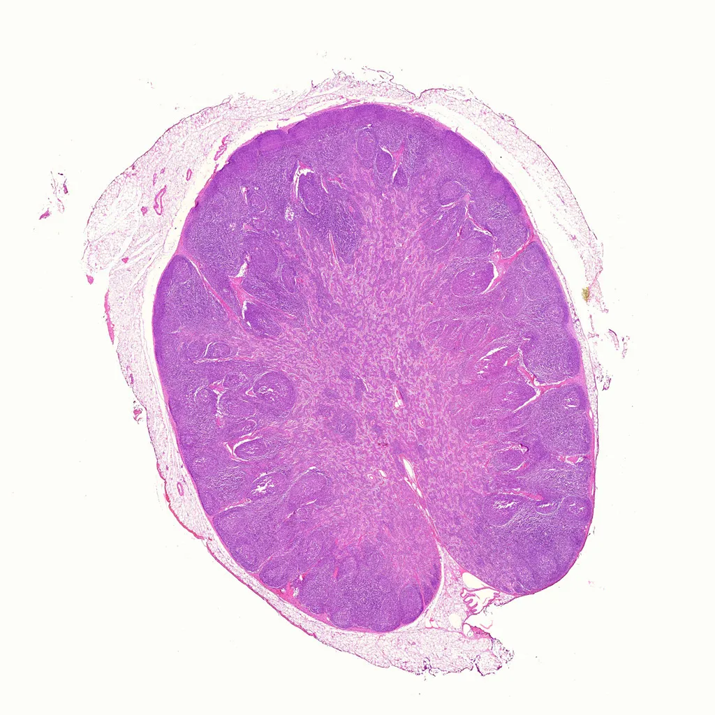
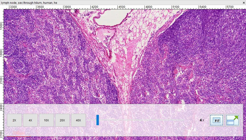
Lymph Node section, H.E. Stain, Prepared Microscope Slides
Original price was: $5.00.$4.00Current price is: $4.00.
Lymph Node section, H.E. Stain, Prepared Microscope Slides
lymphoid follicle histology, germinal center microscopy
H&E stained immune tissue, medical histology lab specimens
Digital file available
Lymph Node section, H.E. Stain, Prepared Microscope Slides
Our Lymph Node slides are expertly sectioned and stained to highlight the distinct functional zones of lymphoid tissue. This specimen is essential for students of histology, immunology, and medicine to understand the structural basis of lymphatic filtration and immune cell activation.
Product Specifications:
-
Dimensions: 25.4mm x 76.2mm (1″ x 3″)
-
Staining: Professionally stained with Hematoxylin and Eosin (H&E). The dense populations of lymphocytes stain a deep purple (basophilic), while the connective tissue framework and vascular elements stain pink/red (eosinophilic).
-
Section Type: Thin section (typically 5–7 microns) providing a complete view of the node from capsule to hilum.
Key Histological Features:
-
Capsule: The outer layer of dense irregular connective tissue that protects the node.
-
Cortex (Outer Zone): Contains highly organized Lymphoid Follicles (Nodules). Look for Germinal Centers, the lighter-staining central areas where B-lymphocytes are actively proliferating.
-
Paracortex: The zone between the cortex and medulla, primarily populated by T-lymphocytes.
-
Medulla (Inner Zone): Organized into Medullary Cords (masses of lymphoid tissue) and Medullary Sinuses (vessel-like spaces) through which lymph flows.
-
Trabeculae: Connective tissue extensions from the capsule that provide structural internal support.
-
Hilum: The concave region where blood vessels enter/exit and efferent lymphatic vessels carry filtered lymph away.
Biological Significance:
-
Immune Filtration: Lymph nodes capture pathogens and cellular debris from the lymph fluid before it returns to the blood circulation.
-
Antigen Presentation: These organs serve as the primary meeting ground where immune cells (like dendritic cells) present “captured” antigens to T-cells and B-cells.
-
Clinical Diagnostic: Changes in lymph node histology (such as follicular hyperplasia) are key indicators of infection, inflammation, or metastatic cancer.
Reference & Further Reading:
For a detailed guide on the microanatomy of lymphoid organs, visit: Lymph node – Wikipedia
Other fixed kits:
Payment & Ordering
Ordering from us is simple and secure. We offer flexible payment options to suit your needs:
-
Online Orders: We primarily accept PayPal (Credit Card payments are supported via PayPal).
-
Alternative Methods: For high-volume orders or regional preferences, we also support Bank Transfer (T/T), Western Union, MoneyGram, WeChat Pay, and Alipay.
-
How to Order: Simply browse our catalog and checkout online, or [Contact Us] for a proforma invoice if using alternative payment methods.

Shipping & Global Delivery
We provide reliable worldwide shipping to ensure your laboratory supplies arrive safely and on time:
-
Fast Processing: Standard orders are processed and shipped within 3 business days.
-
Global Couriers: We partner with leading carriers, including DHL, FedEx, UPS, and official Postal services to offer both express and economy options.
-
Real-time Updates: If your order requires extra preparation time, our team will proactively contact you with a confirmed shipping schedule and tracking details.

Customization & OEM Services
Can’t find a specific specimen? As a leading manufacturer of prepared microscope slides, we specialize in bespoke solutions:
-
Custom Quotations: Send us your specific list, and our technical team will provide a detailed quote within 12 hours.
-
OEM & Private Label: We offer professional OEM services to create customized slide sets tailored to your curriculum or branding requirements.
-
Bulk Inquiries: From rare biological sections to large-scale educational sets, we have the expertise to deliver exactly what you need.
Key Terminology for Microscope Slides
To help you select the most suitable specimen for your research or teaching, please refer to our standard sectioning abbreviations:
-
W.M. (Whole Mount): The entire organism or structure is preserved and mounted on the slide.
-
C.S. (Cross Section): A thin transverse section through the organism or tissue plane.
-
L.S. (Longitudinal Section): A vertical section along the longest plane of the organism.
-
C.S. & L.S.: Both Cross and Longitudinal sections are mounted on the same slide for side-by-side comparison.
Extensive Catalog: Over 3,000+ Prepared Slides
As a leading Microscope Prepared Slides Manufacturer from China, we provide a comprehensive range of biological specimens across multiple disciplines:
| Product Category | Variety Count | Product Category | Variety Count |
| 🌿 Botany | 868 Kinds | 🧬 Cell Biology & Genetics | 64 Kinds |
| 🔬 Histology | 375 Kinds | 🧪 Botany Pathology | 373 Kinds |
| 🦁 Zoology | 459 Kinds | ➗ Mitosis & Meiosis | 66 Kinds |
| 🐾 Animal Pathology | 150 Kinds | 🐣 Embryology | 75 Kinds |
| 🧫 Microbiology | 100 Kinds | 🦟 Parasitology | 120 Kinds |
| 🪨 Petrology (Rock Sections) | 291 Kinds | 🛠️ Customized Sets | Available |
Looking for Custom Microscope Slides?
We specialize in customized preparation to meet specific laboratory or educational requirements.
If your required items are not listed in our standard catalog, we’ve got you covered!
-
Fast Quotation: Email us your specific list, and receive a formal quote within 12 hours.
-
Global Support: Our technical team is ready to assist with your scientific discoveries.
Contact Us Today:
📧 Email: [email protected]
🟢 WhatsApp: +86 18838161683
❓ For more information, please visit our [FAQ Page].
| Weight | 0.1 kg |
|---|---|
| Dimensions | 15 × 20 × 10 cm |
Related products
-
 Histology Slides
Decalcified Compact bone l.s. & c.s. show Osteon and Haversian System, H.E. Stain
$5.00 Add to cart -
-
Sale!
 Histology Slides
1pcs Loose connective tissue (Areolar connective tissue), wholesale prepared microscope slides
Original price was: $3.00.$2.00Current price is: $2.00. Add to cart -
Sale!
 Histology Slides
1pcs mast cells prepared slides, trypan bule injected subcutaneous tissue
Original price was: $3.00.$2.00Current price is: $2.00. Add to cart